
Alopecia / Pattern Baldness Study
Need Help for Alopecia / Pattern Baldness Study!
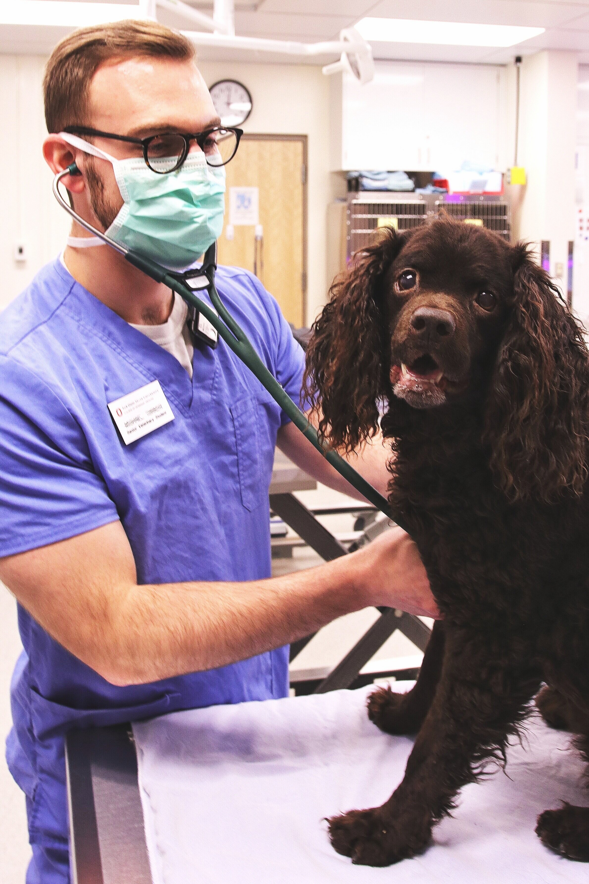
The Alopecia/Pattern Baldness Study for American Water Spaniels is ongoing. The Committee still needs your help! The National presents a great opportunity for us to work together to keep this project moving. For example, we were able to collect blood samples and take photos of 58 dogs while at a recent National. Most of these are unaffected (don't have obvious alopecia/areas of baldness). The geneticist banks the DNA from the blood samples. The researchers want additional AFFECTED dogs as well as the non-affected dogs.
Volunteer your AWS! Your support is needed for the American Water Spaniel genetic research! Please read on, and participate if you qualify.
What is Alopecia?
Alopecia or hair loss is a relatively common finding in American Water Spaniels. Affected dogs tend to show hair loss at a young age (6 months – 1 year), and the hair loss is not associated with infection, itch or other condition. The affected skin may appear dark but, other than that it looks completely normal. The hair loss is typically located on the back legs, the tail, or the neck, around the eyes, but can be located anywhere on the body.
Who is working on this study?
Drs. Brian Husbands, Sheila Torres, and Steven Friedenberg are investigating the genetic basis of hair loss in American Water Spaniels. The goal is to find the genetic mutation associated with the disease so that a test can be developed to decrease the frequency of this disease within the breed. We are soliciting to enroll dogs affected with alopecia/hair loss as well as unaffected/normal dogs.
Information needed to participate:
AKC Registration Number, copy of pedigree, your dog’s medical record, specific photos, and signed consent to join the study.
AWS Pictures Consent to Participate
To learn more about our project, feel free to contact Dr. Brian Husbands at bdhusbands@gmail.com.
AWS DNA Sample Collection Campaign
The Blood Collection Clinics at National are a huge success! Nearly every dog attending and those from surrounding areas donate blood for future research at the free clinics. The samples are sent to the Orthopedic Foundation for Animals to be prepared for future use. The OFA Canine Health Information Center encourages owners donating samples to provide updates regarding any significant changes in the dog's health over its life, especially heritable components. This gesture maximizes the potential usefulness of the sample submitted.
The sample collection campaign is ongoing! The H & G Committee will continue to focus on reaching out to members and non-members - all American Water Spaniel owners - who were not able to attend the National. Breeders are encouraged to contact their puppy clients about this important event!
Blood Collection kits are available from the OFA. The kit (one for each dog you will have sampled) should be taken to your veterinarian to obtain the sample. It is then forward to the CHIC DNA Bank. The H & G Committee will reimburse to the dog owner on communication from the dog's veterinarian that the sample has been taken and forwarded to the OFA.
Frequently Asked Questions About the DNA Bank
Contacts: Aaron Field (aaronfield0@gmail.com); Brian Husbands (bdhusbands@gmail.com; Lois McCracken (lmccracken@ftc-i.net)
Donor Advised Funding For the Canine Health Foundation
AWSC Supports Hemangiosarcoma Research
Hemangiosarcoma - Oncology
02759: Reprogramming the Tumor Immune Niche in Canine Hemangiosarcoma
Grant Status: Open
Grant Amount: $150,000
Jong Hyuk Kim, DVM, PhD; University of Minnesota
July 1, 2020 - June 30, 2022
Sponsor(s): Nespola Chartiable Foundation, Portuguese Water Dog Foundation, Vizsla Club of America Welfare Foundation
Breed(s): -All Dogs
Research Program Area: Oncology - Hemangiosarcoma
Donate to Support this Research Program Area
ABSTRACT
Hemangiosarcoma (HSA) is a common, devastating disease of dogs. The malignant tumor is seen frequently in older Golden Retrievers, German Shepherd Dogs, Portuguese Water Dogs, Labrador Retrievers, and Schnauzers, but it can occur in any dog of any breed at any age. Survival times of dogs with the tumor are short, even with surgical removal and standard of care treatment. Inflammation within the tumor tissue is common in canine HSA, and the immune response may contribute to tumor heterogeneity and prognosis for the dog. Yet, the immunological features in the context of the HSA niche are virtually unknown. The investigators have found that HSA cells have a strong capacity to promote proliferation and differentiation of hematopoietic stem and progenitor cells, with increased inflammatory cytokines, suggesting a niche regulatory function of HSA cells. This study will focus on understanding the functional relationships between HSA cells and immune cells that contribute to the tumor niche to identify molecular mechanisms that regulate critical signaling pathways in canine HSA. This approach will improve our understanding of the tumor immunity and heterogeneity, as well as aid in patient selection for novel immunotherapies.
PUBLICATION(S)
None at this time.
Related Grants
2025: Growth Signaling Pathways in the Pathogenesis and Treatment of Canine Cancer
822: Canine Comparative Oncology and Genomics Consortium (CCOGC)
Study Abstracts of Interest
Alopecia
Title: Alopecia Areata in a Dog: Clinical, Dermoscopic and Histological Features
Citation: Scarampella, F., & Roccabianca, P. (2018). Alopecia Areata in a Dog: Clinical, Dermoscopic and Histological Features. Skin appendage disorders, 4(2), 112–117. https://doi.org/10.1159/000479781
Abstract: Alopecia areata (AA)-like disease is characterized by multifocal patchy hair loss in humans, rodents, dogs, and horses. Remarkable similarities between human and nonhuman AA cases have been reported in terms of clinical presentation, histology, and immune mechanisms of the disease. Canine AA-like lesions most often consist of well-demarcated alopecic patches, frequently but not only involving the face and the head, which extend to the ear pinnae and legs. In some cases, hair loss can have a more generalized distribution. As in humans, hair regrowth is most commonly spontaneous in canine AA-like disease and the resistant cases usually respond to glucocorticoids or cyclosporine treatment. Diagnosis of AA in veterinary medicine relies on presentation, histopathology, and immunohistochemistry and on regrowth following therapy. This case report describes the first dermoscopic evaluation of AA-like disease in a dog with a clinical presentation of symmetrical hair loss.
The takeaway: Studies show similarities between human and canine like alopecia areata (AA) diseases, illustrating that hair regrowth is most commonly spontaneous. However, resistant cases can respond to glucocorticoid or cyclosporine treatment with diagnosis by a veterinary which relies on presentation, histopathology, and immunohistochemistry.
Title: Color-dilution alopecia in dogs.
Citation: Kim, J. H., Kang, K. I., Sohn, H. J., Woo, G. H., Jean, Y. H., & Hwang, E. K. (2005). Color-dilution alopecia in dogs. Journal of Veterinary Science, 6(3), 259–261.
Abstract: Color-dilution alopecia is a relatively uncommon hereditary skin disease seen in "Blue" and other color-diluted dogs. This syndrome is associated with a color-dilution gene. The initial clinical signs are the gradual onset of a dry, dull and poor hair coat quality. Hair shafts and hair regrowth are poor, and follicular papules may develop and progress to frank comedones. Hair loss and comedo formation are usually most severe on the trunk, especially color-diluted area on the skin. Six cases of color-dilution alopecia are reported in 3 months to 10 years old dogs. The breeds of dogs are blue Doberman Pinscher, Miniature Pinscher, Dachshund, and Schnauzer. Grossly, extensive partial hair loss was seen on the skin. Histopathologically, the epidermis is relatively normal but may be hyperplastic. Hair follicles are characterized by atrophy and distortion. Heavily clumped melanin is present in the epidermis, dermis and hair follicles.
The takeaway: A version of alopecia involves color-dilution seen most commonly in blue or other color diluted dogs such as Doberman Pinschers, Miniature Pinscher, Dachshund, and Schnauzers. Studies show that the hair follicles can be characterized by atrophy, meaning the follicles are degrading, with melanin heavily clumped in those areas.
Title: Estrogen-related alopecia due to polycystic ovaries in a terrier dog
Citation: Selk Ghaffari, M., Dezfoulian, O., Aldavood, S.J. et al. Estrogen-related alopecia due to polycystic ovaries in a terrier dog. Comp Clin Pathol 18, 341–343 (2009). https://doi-org.proxy.bsu.edu/10.1007/s00580-009-0815-x
Abstract: Sex hormone-related alopecia is a rare clinical condition in dogs. A 9-year-old female dog was presented with a history of symmetrical alopecia on the caudal aspects of both thighs. A dermatophyte culture, skin scrapes, and acetate strip examinations were negative for dermatophytes, parasites, and yeasts. The only abnormality in abdominal ultrasonography was multiple cystic follicles within the ovaries. This increased the possibility of hyperestrogenism due to ovarian cysts. Serum estrogen assay indicated elevated estrogen concentration. Ovariohystorectomy was performed and tissue samples of uterus, ovaries, and skin biopsies were submitted for histopathological examination. In histological examination, the polycystic ovary was characterized by multiple follicular cysts. Results of histopathological findings in skin biopsies were similar to that reported with hyperestrogenism. In view of information achieved from the presented case, hyperestrogenism should be included in the differential diagnosis for dogs with clinical manifestation of symmetrical alopecia.
The takeaway: This study illustrated that alopecia could be linked with estrogen related to polycystic ovaries. The alopecia was found symmetrically on both thighs, with cystic follicles found within the ovaries as the only abnormality. Estrogen was found in concentration, and should be included as a differential diagnosis for canines with alopecia.
Neutering
Title: Neutering dogs: effects on joint disorders and cancers in golden retrievers.
Citation: Torres de la Riva, G., Hart, B. L., Farver, T. B., Oberbauer, A. M., Messam, L. L., Willits, N., & Hart, L. A. (2013). Neutering dogs: Effects on joint disorders and cancers in golden retrievers. PLoS ONE, 8(2). doi:10.1371/journal.pone.0055937
Abstract: In contrast to European countries, the overwhelming majority of dogs in the U.S. are neutered (including spaying), usually done before one year of age. Given the importance of gonadal hormones in growth and development, this cultural contrast invites an analysis of the multiple organ systems that may be adversely affected by neutering. Using a single breed-specific dataset, the objective was to examine the variables of gender and age at the time of neutering versus leaving dogs gonadally intact, on all diseases occurring with sufficient frequency for statistical analyses. Given its popularity and vulnerability to various cancers and joint disorders, the Golden Retriever was chosen for this study. Veterinary hospital records of 759 client-owned, intact and neutered female and male dogs, 1-8 years old, were examined for diagnoses of hip dysplasia (HD), cranial cruciate ligament tear (CCL), lymphosarcoma (LSA), hemangiosarcoma (HSA), and mast cell tumor (MCT). Patients were classified as intact, or neutered early (<12 mo) or late (≥12 mo). Statistical analyses involved survival analyses and incidence rate comparisons. Outcomes at the 5 percent level of significance are reported. Of early-neutered males, 10 percent were diagnosed with HD, double the occurrence in intact males. There were no cases of CCL diagnosed in intact males or females, but in early-neutered males and females the occurrences were 5 percent and 8 percent, respectively. Almost 10 percent of early-neutered males were diagnosed with LSA, 3 times more than intact males. The percentage of HSA cases in late-neutered females (about 8 percent) was 4 times more than intact and early-neutered females. There were no cases of MCT in intact females, but the occurrence was nearly 6 percent in late-neutered females. The results have health implications for Golden Retriever companion and service dogs, and for oncologists using dogs as models of cancers that occur in humans.
The takeaway: A comparison of intact, early neutering, and late neutering of Golden Retrievers showed that twice as many early neutered males were diagnosed with hip dysplasia as compared to intact males. No intact dogs were diagnosed with cranial cruciate ligament tear compared to 5% (females) and 8% (males) of early neuter dogs. Early neutered males were diagnosed 3 times more than intact males with lymphosarcoma, however hemangiosarcoma was diagnosed 4 times in late neuter females than intact and early neutered females.
Title: Long-term health effects of neutering dogs: comparison of Labrador Retrievers with Golden Retrievers.
Citation: Hart, B. L., Hart, L. A., Thigpen, A. P., & Willits, N. H. (2014). Long-Term health effects of Neutering Dogs: Comparison of Labrador retrievers with Golden Retrievers. PLoS ONE, 9(7). doi:10.1371/journal.pone.0102241
Abstract: Our recent study on the effects of neutering (including spaying) in Golden Retrievers in markedly increasing the incidence of two joint disorders and three cancers prompted this study and a comparison of Golden and Labrador Retrievers. Veterinary hospital records were examined over a 13-year period for the effects of neutering during specified age ranges: before 6 mo., and during 6-11 mo., year 1 or years 2 through 8. The joint disorders examined were hip dysplasia, cranial cruciate ligament tear and elbow dysplasia. The cancers examined were lymphosarcoma, hemangiosarcoma, mast cell tumor, and mammary cancer. The results for the Golden Retriever were similar to the previous study, but there were notable differences between breeds. In Labrador Retrievers, where about 5 percent of gonadally intact males and females had one or more joint disorders, neutering at <6 mo. doubled the incidence of one or more joint disorders in both sexes. In male and female Golden Retrievers, with the same 5 percent rate of joint disorders in intact dogs, neutering at <6 mo. increased the incidence of a joint disorder to 4-5 times that of intact dogs. The incidence of one or more cancers in female Labrador Retrievers increased slightly above the 3 percent level of intact females with neutering. In contrast, in female Golden Retrievers, with the same 3 percent rate of one or more cancers in intact females, neutering at all periods through 8 years of age increased the rate of at least one of the cancers by 3-4 times. In male Golden and Labrador Retrievers neutering had relatively minor effects in increasing the occurrence of cancers. Comparisons of cancers in the two breeds suggest that the occurrence of cancers in female Golden Retrievers is a reflection of particular vulnerability to gonadal hormone removal.
The takeaway: Both breeds, Golden Retriever and Labrador Retriever showed no notable difference between them with both breeds showing twice the incidence of joint disorders in early neutering compared to intact. The study also found that neutering at all periods in life through 8 years of age increased the rate of cancer by 3 to 4 times compared to intact.
Title: Neutering is associated with developing hemangiosarcoma in dogs in the Veterinary Medical Database: An age and time-period matched case-control study (1964–2003)
Citation: Robinson, K. L., Bryan, M. E., Atkinson, E. S., Keeler, M. R., Hahn, A. W., & Bryan, J. N. (2020). Neutering is associated with developing hemangiosarcoma in dogs in the Veterinary Medical Database: An age and time-period matched case-control study (1964-2003). The Canadian veterinary journal = La revue veterinaire canadienne, 61(5), 499–504.
Abstract: The hypothesis that neutered dogs in the Veterinary Medical Database (VMDB) are at increased risk for developing hemangiosarcoma (HSA) was tested. Dogs (n = 5736) were diagnosed with HSA from a population of 2 106 324 dogs in the VMDB from 1964 to 2003. A case-control design matched on age and time period was created for general, cardiac, and splenic HSAs. A logistic regression analysis was performed including breed. Spayed females had an odds ratio (OR) of 1.59 for splenic, 1.47 for cardiac, and 1.72 for HSA in general. Castrated males had an OR of 1.26 for splenic and 1.14 for HSA in general compared to intact males. Controlled for historical time period and patient age, VMDB data support that neutering is associated with development of splenic HSA and HSA in general in both male and female dogs, but not cardiac HSA with an apparently lower than previously described magnitude of association.
Key clinical message:
This case-control design confirms an association between neutering and development of HSA and splenic HSA, but not cardiac HSA, in both male and female dogs. By controlling for time period at diagnosis, the bias of recent early neuter practices is eliminated, suggesting early neuter is not a principal driver of this effect.
Confidential Health Survey
for the
American Water Spaniel
The Health and Genetics Committee is so excited to be able to make an on-line Health Survey available to all owners of American Water Spaniels. The results of this confidential survey will provide insight into the overall health of our breed. The survey is available through the website of the Orthopedic Foundation for Animals (OFA) a name we all know and trust. All entries and results are completely confidential. No names are recorded, no stamps are needed and everyone can see the results for the overall entries instantly. OFA is providing this service to breed clubs at no cost. The survey has a very inclusive set of health conditions which have been known to occur in dogs. You will take one survey for each dog. When you start, you will see a concise list of conditions which may have affected your dog, such as cancer/tumors, eye disorders or liver disorders. When you select one of these, the next step will offer you a comprehensive listing within that category. If you don’t see a diagnosis that your dog may have received from a veterinarian, you should select “Other” and then you will be able to describe that diagnosis. Please take a few moments of your time to fill out the survey for as many of your dogs as possible. All living or deceased dogs are needed, healthy included, so that a true picture of breed health can be seen. The survey asks that your results be based on a diagnosis from your veterinarian, so you may want to review the survey first and check your dog’s health records for accuracy. The results of this survey will assist the club in observing the overall health picture of AWS. We will be able to make better decisions about where to focus our efforts and money. The background statistics will allow us to see if there are trends or new concerns over the coming years and will help us garner attention and research funds from the AKC Canine Health Foundation (CHF) if needed. Educational opportunities for breeders and owners can be planned if we find breed-specific health issues. Your efforts to complete this survey will be more appreciated than we can say. Please pass the word to all AWS owners you know. AWS is a breed of small numbers and we need to do all we can to keep them healthy!
Please click below for direct access to survey:
